Introduction
An antimicrobial is an agent that
kills or inhibits the growth of microorganisms. The microbial agent may be a
chemical compounds and physical agents. These agents interfere with the growth
and reproduction of causative organisms like bacteria, fungi, parasites, virus
etc. Antimicrobial substances
can be produced by certain group or species of bacteria with the capacity to
inhibit the growth of pathogenic and spoilage microorganisms. For instance, the
microbial compounds include organic acids, hydrogen peroxide, diacetyl and
bacteriocins. The use of these substances with antimicrobial properties is
known to have been common practice for at least 2000 years. Antimicrobial
agents are widely used in food system and resistance management which plays a
role in controlling both pathogenic and spoilage microbe to grow. Nowadays,
consumers demand “natural” and “minimally processed” food, the interest in
naturally produced antimicrobial agents such as bacteriocins is on the rise of
demand. The discovery of bacteriocins gave a new way for food development in
better hygienic quality.
Bacteriocins
are a kind of ribosomal synthesized antimicrobial peptides produced by
bacteria, which can kill or inhibit bacterial strains closely-related or
non-related to produced bacteria, but will not harm the bacteria themselves by
specific immunity proteins. Bacteriocins become one of the weapons against
microorganisms due to the specific characteristics of large diversity of
structure and function, natural resource, and being stable to heat.
Bacteriocins are categorized in several ways, including producing strain,
common resistance mechanisms, and mechanism of killing.
Bacteriocins are categorized in several ways,
including producing strain, common resistance mechanisms, and mechanism of
killing. Moreover, bacteriocins can be found in numerous Gram-positive and
Gram-negative bacteria, those produced by lactic acid bacteria (LAB) have
received special attention in recent years due to their potential application
in the food industry as natural biopreservatives.
LAB
also known as Lactobacillalesare either rod-shaped (bacillus), or spherical
(coccus).Different classes of LAB bacteriocins have been identified on the
basis of biochemical and genetic characterization. These bacteriocins have been
reported to inhibit the growth of Listeria monocytogenes, Staphylococcus
aureus, Enterococcus faecalis and Clostridium tyrobutyricum. In this experiment, Salmonella bacteria and
Escherichia coli are used.
Objective
To determine the antimicrobial effects
of extracelluar extracts of selected LAB strains
Materials
and reagents
MRS broth
Sterile filter paper disk (50mm x 50mm)
Forceps
Sterile universal bottles
Cultures of LAB and spoilage/ pathogenic organisms
(Escherichia coli and Salmonella )
Bench-top refrigerated centrifuge
Incubator 30°C and 37°C
UV/Vis spectrophotometer
Distilled deionized water
Trypticase soy agar
Brain heart infusion agar
Yeast extract
Bunsen burner
Pipette and tips
Sterile petri dish
96 well plate
Procedure
Part 1: Determination of bacteriocin
activity via agar diffusion test
1. All the petri dishes are labelled
according to the spoilage organisms and strains of LAB used.
2. Each plate are used for one strain of
spoilage organism and one strain of LAB by dividing the plate into 2 sides,
each side for one replicate.
3. 2 strains of LAB and 2 strains of
spoilage/ pathogenic organisms are given to each group.
4. 10 ml of trypticase soy-yeast extract
agar (TSAYE) are loaded into the labelled petri dishes using pipette and “
figure of 8 ” is performed to ensure that the entire surface of the plate is
covered by the agar. The petri dishes were left aside for the agar to be
solidified.
5. 2 ml of the broth containing the
spoilage organism are innoculated into 10 ml of brain heart infusion (BHI) agar
and the misture are vortexed.
6. The mixture is then quickly loaded on
top of the TSAYE agar layer, it is ensured to cover the entire surface and the
petri dishes are left aside for the mixture to solidify.
7. The broth containing LAB cultures are
centrifuged and the supernatant obtained are used as the extracellular extracts
by draining off the excess extract.
8. A sterile filter paper disk is picked
up aseptically using sterile forceps and the disk is dipped into the
extracellular extract.
Figure
1: A sterile filter paper disk is picked up aseptically using sterile forceps

Figure
2: The disk is dipped into the extracellular extract.

9. The
paper disk is placed on top of the solidified BHI agar.
Figure
3: The disk are placed on top of the agar.

10. The
plates are incubated for 24-48 hours at 37°C.
Figure
4: The plates are placed into incubator.

11. The inhibition zones (in cm) is measured after incubation and the readings are recorded.
Part 2: Determination of bacteriocin
activity via optical density
1. The broth containing LAB cultures are
centrifuged. The supernatant are used as the extracellular extracts.
2. Each group are given 2 strains of LAB
and 2 strains of spoilage/pathogenic organisms.
3. 10 µl of double-strength MRS are added
with 10 µl of cultures containing spoilage/ pathogenic organism and the mixture
is vortexed.
4. A serial dilution of LAB extracellular extract with
MRS was prepared with final volume of 1000 μl in each column. The volume of the
LAB extracellular extract and MRS needed for each column is shown as below:
Table 1: Serial dilution
|
Mixture
|
Dilution
|
|||||
|
0x
|
2x
|
10x
|
50x
|
100x
|
Control
|
|
|
LAB Extracellular
Extract (μl)
|
1000
|
500
|
200
|
200
|
500
|
0
|
|
MRS Broth (μl)
|
0
|
500
|
800
|
800
|
500
|
1000
|
|
Total (μl
|
1000
|
1000
|
1000
|
1000
|
1000
|
1000
|
Figure 5: LAB extracellular
extract is loaded according to the volume required.

Figure
6: MRS broth is added to the LAB extracellulat extract.

Figure
7: Serial dilution is done.

5. 50 μl of each extracellular extracts dilution are added
into mixture as prepared in step 3.
Figure 8: The extracellular
extracts dilution are loaded into 96 well plate.

6. The mixtures are incubated for 12-15 hours at 370C.
7. A control
is prepared using 5 ml of
double-strength MRS, 1 ml of cultures containing spoilage/pathogenic bacteria
and 10 ml of MRS. The mixtures are incubated for 12-15 hours at 370C.
8. A
negative-control is prepared for “auto-zero” via spectrophotometer. 5 ml of
double-strength MRS are added to the control.
Figure 9: Spectrophotometer

9. Upon incubation, the optical density of the spoilage/pathogenic bacteria are measured at 600 nm. The same method is performed for the control as well.
10. One arbitrary
unit (AU) is defined as the dilution factor of the extracellular extract that
inhibited 50% of the spoilage/pathogenic bacteria growth and expressed as
AU/ml.
11. 50% of the spoilage/pathogenic bacteria growth
are determined from the OD600 of the control.
Results
Part I: Determination of bacteriocin
activity via agar diffusion test
Figure 10: Petri dish containing LAB 1 and E.coli

Figure 11: Petri dish containing LAB 1 and Salmonella
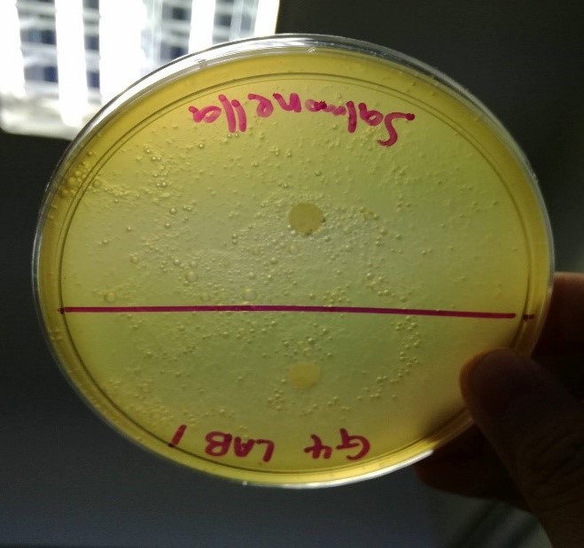
Figure 12: Petri dish containing LAB 2 and E.coli

Figure 13: Petri dish containing LAB 2 and Salmonella

Table 1: Inhibition zone of strains of LAB on strains
of spoilage/pathogenic bacteria
Strains of LAB
|
Strains of
spoilage/ pathogenic bacteria
|
Inhibition zone
(cm)
|
LAB 1
|
E.coli
|
1.0
|
Salmonella
|
0.6
|
|
LAB 2
|
E.coli
|
0.8
|
Salmonella
|
0.6
|
Part
II: Determination of bacteriocin activity via optical density
Serial dilution of extracellular extract
LAB 1
Figure 14: 96 well plate containing E.coli and Salmonella
with different dilutions of LAB 1

Table 2: OD600 of E.coli according to
different dilutions of LAB 1
Dilutions
|
OD600 of spoilage/ pathogenic
bacteria
|
|||
Strain 1: E.coli
|
||||
Reading 1
|
Reading 2
|
Reading 3
|
Average
|
|
0x
|
0.263
|
0.436
|
0.433
|
0.3773
|
2x
|
0.712
|
1.399
|
1.389
|
1.1667
|
10x
|
0.835
|
1.436
|
1.491
|
1.254
|
50x
|
0.792
|
0.963
|
1.415
|
1.0567
|
100x
|
0.582
|
0.819
|
1.102
|
0.834
|
Equation
|
y
= 0.0803x + 0.6967
|
|||
OD600 of control
|
0.582
|
0.819
|
1.102
|
0.834
|
50% of OD600
|
0.291
|
0.410
|
0.551
|
0.417
|
AU/ml
|
X=
(0.417-0.697)/0.0803
= -3.487
|
|||

Graph 1: OD600 of E.coli against different dilutions
of LAB 1
Table 3: OD600 of Salmonella according to
different dilutions of LAB 1
Dilutions
|
OD600 of spoilage/ pathogenic
bacteria
|
|||
Strain 2: Salmonella
|
||||
Reading 1
|
Reading 2
|
Reading 3
|
Average
|
|
0x
|
0.329
|
0.323
|
0.159
|
0.2703
|
2x
|
1.153
|
1.324
|
0.790
|
1.089
|
10x
|
1.468
|
1.476
|
0.961
|
1.3017
|
50x
|
1.428
|
1.340
|
0.920
|
1.2293
|
100x
|
0.956
|
0.997
|
0.681
|
0.878
|
Equation
|
y
= 0.1356x + 0.547
|
|||
OD600 of control
|
0.956
|
0.997
|
0.681
|
0.878
|
50% of OD600
|
0.478
|
0.499
|
0.341
|
0.439
|
AU/ml
|
X=
(0.439-0.547)/0.1356
=-0.796
|
|||

Graph 2: OD600 of Salmonella against
different dilutions of LAB 1
Discussion
Part I: Determination of bacteriocin activity via agar diffusion test
Bacteriocins are protein which is secreted by bacteria to inhibit the growth of other closely- related bacterial strain. Bacteriocin causes the destruction of the membrane potential, forming the pores on the pathogenic bacteria. Bacteriocin also inhibits the protein synthesis of the pathogenic bacteria. Besides that, it inhibits the nucleolytic activity of the pathogenic bacteria strains which breaks down the DNA chromosomes as well as RNA. In this experiment, bacteriocin is produced by lactic acid bacteria (LAB).
Lactic acid bacteria are rod-shaped bacilli or cocci
characterized by an increase tolerance to a lower pH range. The production of
organic acids such as lactic acid and acetic acid by lactic acid bacteria
causes acidification, hence it can also be used to inhibit spoilage bacteria.
Bacteriocin are usually effective against
Gram-positive bacteria. Although Gram-positive bacteria has thick cell wall
made of protein and polysaccharides, but is easily digested by acid produced by
lactic acid bacteria (LAB). LAB may be not efficient enough to inhibit
Gram-negative bacterium because the cell wall is made from lipid layer which
prevent acid from being digested by acid produced by LAB. In this experiment,
the spoilage bacteria that we used are Escherichia
coli and Salmonella,
in which both are Gram-negative bacteria.
In agar diffusion test, a filter-paper disk saturated
with bacteriocin of LAB is placed on the surface of the agar. The compound
diffuses from the filter paper into the agar. The concentration of the compound
near the disk would be the highest, and will decreases as distance from the
disk increases. The
effectiveness of LAB antimicrobials towards the growth of spoilage
microorganisms can be determined by measuring the inhibition zones around the
LAB-staining paper disks. Inhibition zone is the clear region around the paper
disc, which is an indication of the absence or the effective inhibition of
microbial growth by the antimicrobial agent. The larger the inhibition zones,
the higher the degree of sensitivity of spoilage microorganisms to LAB
antimicrobials.
As a result, the average measurement of inhibition
zone for Escherichia coli is 0.9cm while for
measurement of inhibition zone for Salmonella
is 0.6cm. The inhibition effect of LAB is greater towards Escherichia coli
compared to Salmonella.
Part
2: Determination of bacteriocin activity via optical density
Optical density is a measurement of the concentration of bacteria in a suspension, which can be measured using a spectrophotometer. Spectrophotometer is an instrument which can be used to measure the amount of light scattered at a specific wavelength when passes through a medium. When visible light passes through a cell suspension, the light is scattered. Greater degree of scatter indicates that more bacteria are present. A spectrophotometer can be set at a wavelength of 420 – 660 nm. In this experiment, the OD600 is measured. OD600 is an abbreviation indicating the optical density of a sample is measured at a wavelength of 600 nm, which is much preferable because at this wavelength, the cells will not be killed due to the exposure of too much light intensity.
One arbitrary (AU) is known as the dilution factor of
the extracellular extract that inhibited 50% of the spoilage or pathogenic
bacteria growth and expressed as AU/ml.
Control: Abs600 = Z. Thus, 50% of Z = Z/2
y = mx + c; Thus, x = (y-c)/m
When y = Z/2, Thus x = (Z/2 -c)/m
Both graph plotted from the data we obtained has
positive gradient from 0x to 10x dilution, which means LAB 1 shows positive
inhibition on Escherichia coli
and on Salmonella. The lower
the concentration of extracellular extract, the lower concentration of
bacteriocin, the higher the growth rate of bacteria. However, the graphs later
show negative gradient from 10x to 100x dilution. There is a decreasing trend
on the growth of bacteria from 10x to 100x dilution. The result obtained might
not be accurate due to the improper preparation of the serial dilution
solution. This causes the result obtained is not like what it is supposed to
be. This means that both graph should always have positive gradient from 0x to
100x dilution.

Conclusion
Lactic acid bacteria (LAB) is a useful bacterium used
to produce bacteriocin that can inhibit the growth of spoilage bacteria like Escherichia coli and Salmonella.
Reference
No comments:
Post a Comment